… come solo gli Anatomisti sanno fare!
IV Florence International Symposium on Advances in CARDIOMyopathies
Fondazione Menarini – Firenze 9-10 settembre 2021

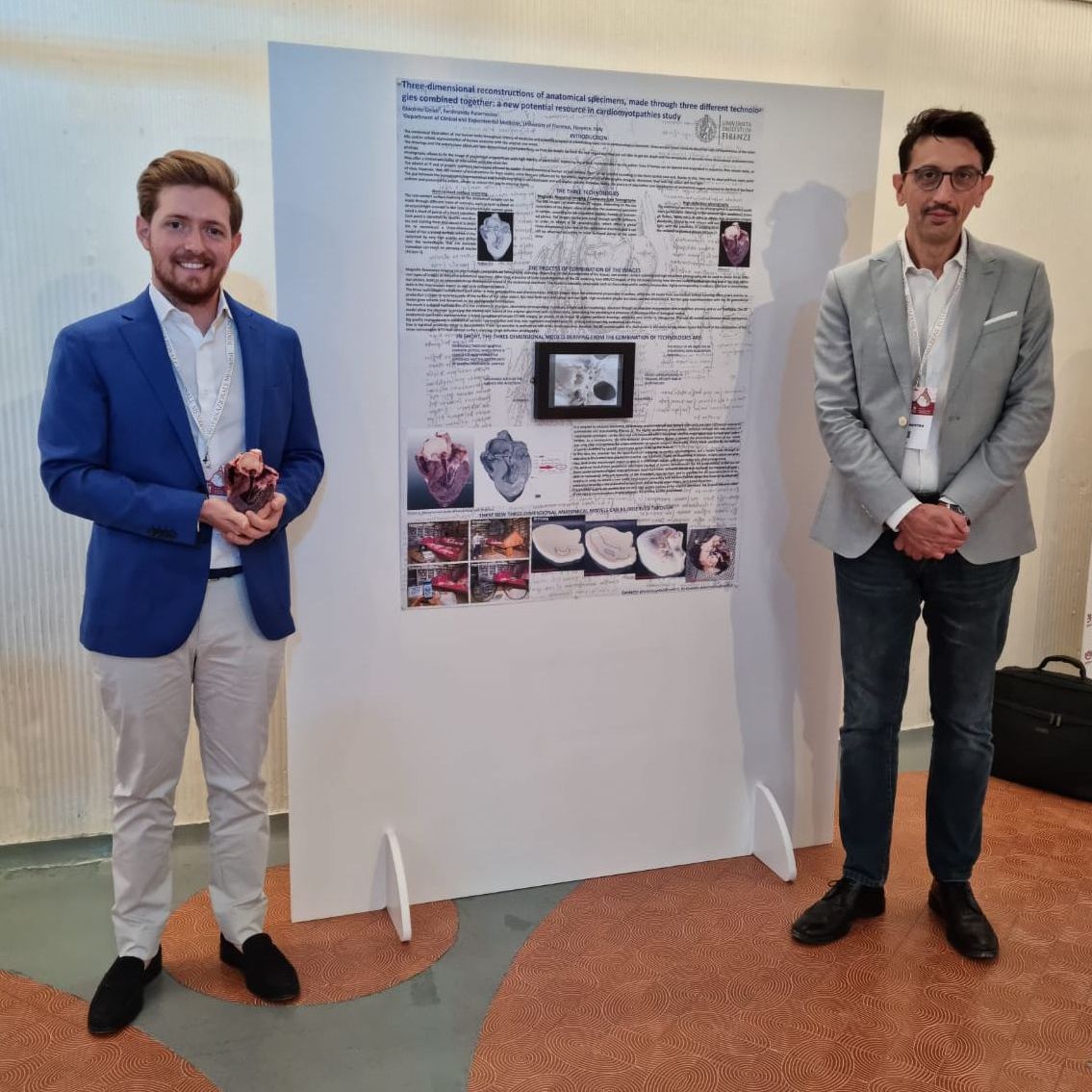
Three-dimensional reconstructions of anatomical specimens, made through three different technologies combined together: a new potential resource in cardiomyopathies study
Giacomo Gelati, Ferdinando Paternostro
Department of Clinical and Experimental Medicine, University of Florence, Florence, Italy
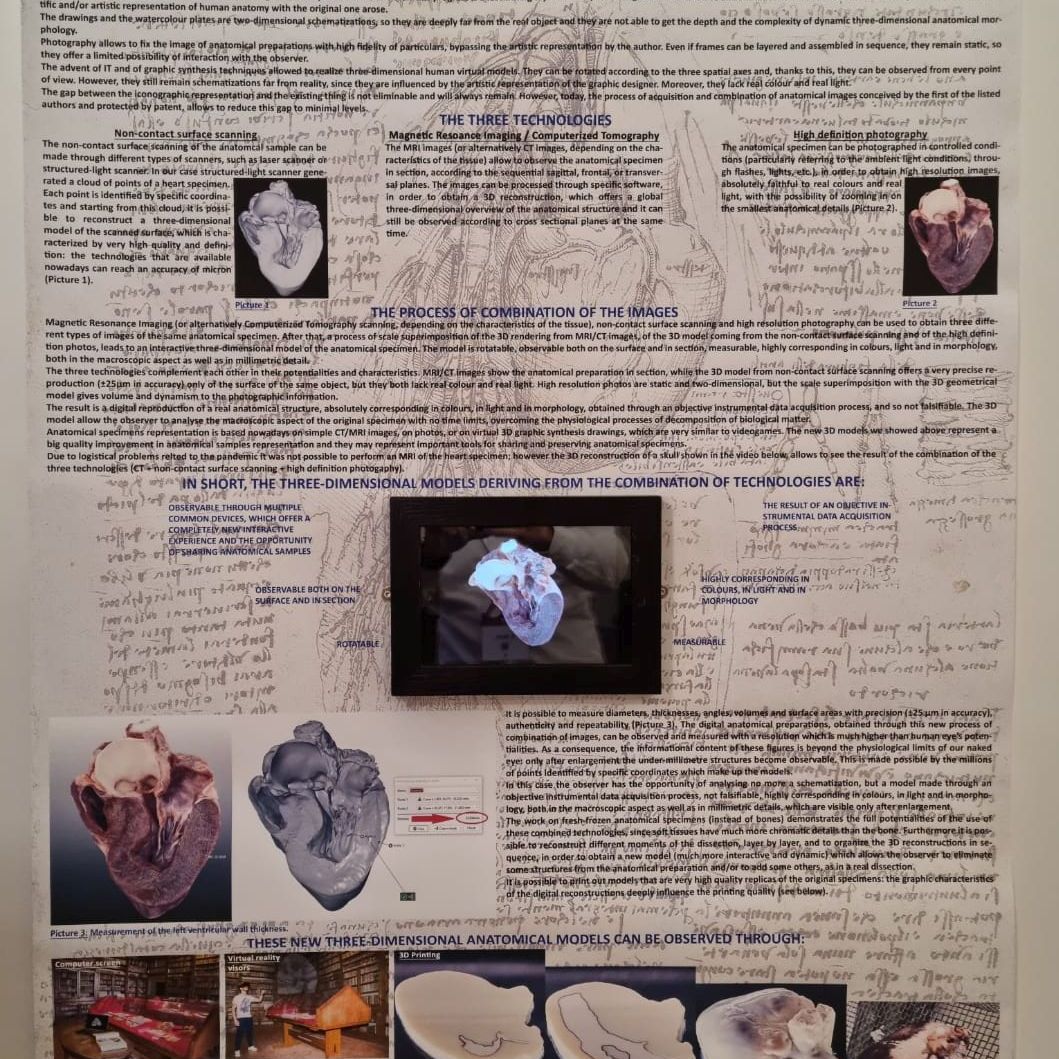
Magnetic Resonance Imaging (or alternatively Computerized Tomography scanning, depending on the characteristics of the tissue), non-contact surface scanning and high resolution photography can be used to obtain three different types of images of the same anatomical specimen. After that, a process of scale superimposition of the 3D rendering from MRI/CT images, of the 3D model coming from the non-contact surface scanning and of the high resolution photos, leads to an interactive three-dimensional model of the anatomical specimen. The model is rotatable, observable both on the surface and in depth, measurable, corresponding in colours, light and in morphology, both in the macroscopic aspect as well as under the millimetre.
The three technologies complement each other in their potentialities and characteristics. MRI/CT images show the anatomical preparation in depth, while the 3D model from non-contact surface scanning offers a very precise reproduction (±25μm in accuracy) just of the surface of the same object, but they both lack real colour and real light. High resolution photos are static and two-dimensional, but the scale superimposition with the 3D geometrical model, gives volume and dynamism to the photographic information.
The result is a digital reproduction of a real anatomical structure, absolutely corresponding in colours, in light and in morphology, obtained through an objective instrumental data acquisition process, and so not falsifiable. The 3D models allow the observer to analyse the macroscopic aspect of the original specimen with no limits of time, overcoming the physiological processes of decomposition of biological matter. It is possible to measure diameters, thicknesses, angles, volumes and surface areas with precision (±25μm in accuracy), authenticity, and repeatability. Printing out the models it is possible to obtain very high quality replicas of the original specimens: the graphic characteristics of the digital reconstructions deeply influence the printing quality.
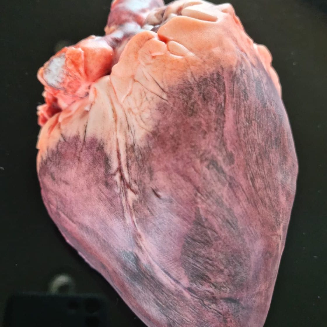
With this work we present the 3D reconstruction of a fresh-frozen human heart (provided by Nicola’s Foundation Onlus at Iclo Teaching and Research Center – Verona, Italy), in order to show the full potential of the models described above, which may represent important tools for sharing and preserving anatomical specimens.
The process of combination of technologies is conceived by the first of the present authors and protected by a patent.