
Jacopo Junio Valerio Branca1, Giulia Guarnieri1, Annamaria Morelli1 , Carlo Benedini2 , Niccolò Fagni 3, Massimo Gulisano1 , Alessandra Pacini1, Ferdinando Paternostro1
- Experimental and Clinical Medicine, University of Firenze, Firenze, ITA
- Physical Medicine and Rehabilitation, ICLO Teaching and Research Center, Verona, ITA
- Otorinolaringoiatry, Azienda Ospedaliero-Universitaria Senese (UOSA), Siena, ITA
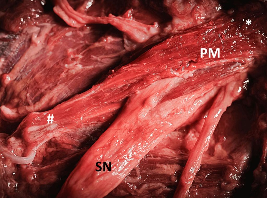
Knowledge of anatomical variability is extremely important in order to better understand the etiology of pain, if present, or to avoid iatrogenic consequences. Sometimes the anatomical “anomalies” have the same anamnesis but different causes. For example, sciatic neuralgia may be caused by a herniated disc or it may have a different origin. The sciatic nerve (SN), also known as the ischial nerve, is the widest in the human body. This huge peripheral nerve originates from the roots of the lumbosacral plexus (L4-S3) and passes through the great sciatic foramen, under the piriformis muscle (PM). However, there is much variability in the pattern of SNs about the muscle, which has been known since the first half of the 20th century. In the present study, we describe six different case reports of anatomical variations of the SN and its interplay with the PM. The observations were made during dissection classes at the ICLO Teaching and Research Centre (Verona, Italy), on both male and female cadavers aged between 58 and 84 years. The SN was reported as a single and divided nerve into the tibial nerve (TN) and the common peroneal nerve (CPN), passing alone above, below, or between the PM. However, the two parts of the SN may also interact with the PM in different ways, adding to the anatomical variability. A thorough knowledge of the anatomical variations in any part of the human body is extremely important. The various techniques used, from imaging to autopsy or surgery, are also useful in the SN pathway. Thus, the anatomical features and the understanding of each variation are useful for a correct approach that can lead to an effective and correct treatment with a favorable outcome.
Acknowledgements
We are extremely grateful to Dr. Alessandro Palazzolo, Dr. Daniele Pignatelli, Dr. Cristiana Veltro, Anna Venzi, and Aurora Baroni for their skills and dedication demonstrated during the dissection classes performed together with the ICLO staff and Prof. Ferdinando Paternostro.
Branca J, Guarnieri G, Morelli A, et al. (May 11, 2024) Sciatic Nerve and Its Anatomical Variations: In-Depth Understanding Acquired During Dissection Classes.
Cureus 16(5): e60083. doi:10.7759/cureus.60083