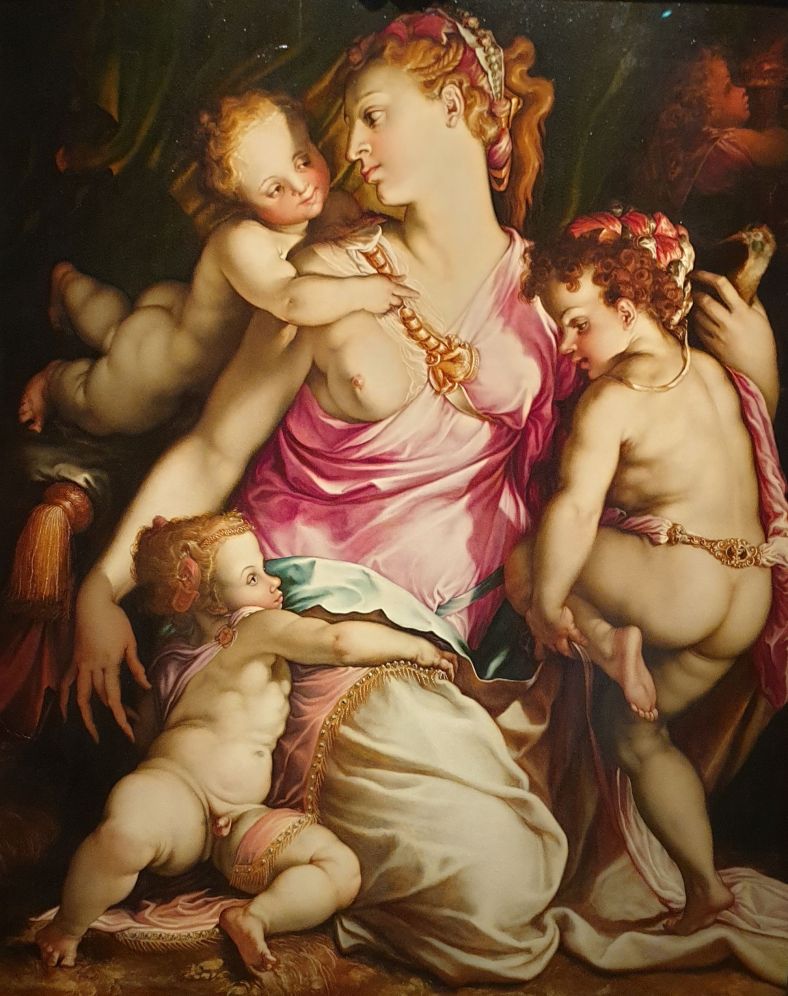
In questo articolo, il dipinto La Carità di Francesco de Rossi, noto come Salviati (1510-1563), viene studiato attraverso una prospettiva multidisciplinare che unisce Anatomia umana, Paleopatologia e Storia dell’Arte. Sebbene non sia possibile formulare una diagnosi definitiva a causa della natura artistica dell’opera, le anomalie anatomiche rappresentate nel dipinto sono esaminate considerando due ipotesi principali: mastite cronica o tumore al seno. Questo studio evidenzia ancora una volta come l’incontro tra Medicina e Arte possa offrire un prezioso contributo sia alla comprensione delle malattie del passato sia all’educazione dei futuri Medici, aiutandoli a sviluppare un più attento occhio clinico.
Paternostro, F., Lippi, D., Zucchini, E., Nori, J., Galassi, F.M., Nerlich, A.G., & Bianucci, R. (2024). Italian Journal of Anatomy and Embryology 128(2): 55-60. https://doi.org/10.36253/ijae-15580
Ferdinando Paternostro, Wei-Jin Hong, Guo-Sheng Zhu, Jeremy B. Green, Milan Milisavljevic, Mikaela V. Cotofana, Michael Alfertshofer, S. Benoit Hendrickx, Sebastian Cotofana https://onlinelibrary.wiley.com/doi/10.1111/jocd.16631
Aesthetic neuromodulator injections of the upper face are frequently performed to temporarily block muscular actions of the periorbital muscles to ultimately reduce skin rhytids. However, the adverse event rate in the literature for toxin-induced blepharoptosis ranges from 0.51% to 5.4%.
To identify access pathways by which injected neuromodulator product can travel from extra-to intra-orbital and therefore affect the levator palpebrae superioris muscle.
Nine non-embalmed human body donors were investigated in this study with a mean age at death of 72.8 (16.1) years. The 18 supraorbital regions were injected in 28 times (14 for supratrochlear and 14 for supraorbital) with 0.5 cc, whereas eight cases (four for supratrochlear and four supraorbital) were injected with 0.1 cc of colored product. Anatomic dissections were conducted to identify structures stained by the injected color.
The results of this injection-and dissection-based study revealed that both the supratrochlear and the supraorbital neurovascular bundles are access pathways for injected neuromodulator products to reach the intra-orbital space and affect the levator palpebrea superioris muscle. Out of 36 conducted injection passes, seven (19.44%) resulted in affection of the sole elevator of the eyelid of which 100% occurred only at an injection volume of 0.5 cc and not at 0.1 cc.
Clinically, the results indicate that a low injection volume, a superficial injection for the supraorbital location, and angling the needle tip away from the supratrochlear foramen (toward the contralateral temple) when targeting the corrugator supercilii muscles, can increase the safety profile of an aesthetic toxin glabellar treatment.
Paternostro, F., Hong, J., Zhu, S., Green, J. B., Milisavljevic, M., Cotofana, M. V., Alfertshofer, M., Hendrickx, S. B., & Cotofana, S. Simulating Upper Eyelid Ptosis During Neuromodulator Injections—An Exploratory Injection and Dissection Study. Journal of Cosmetic Dermatology. https://doi.org/10.1111/jocd.16631
Francesco P. Bernardini, Brent Skippen, Raul Cetto, Mariana Calomeni, Sebastian Cotofana, Simone Ugo Urso, Ferdinando Paternostro, Morris E. Hartstein Journal of Cosmetic Dermatology
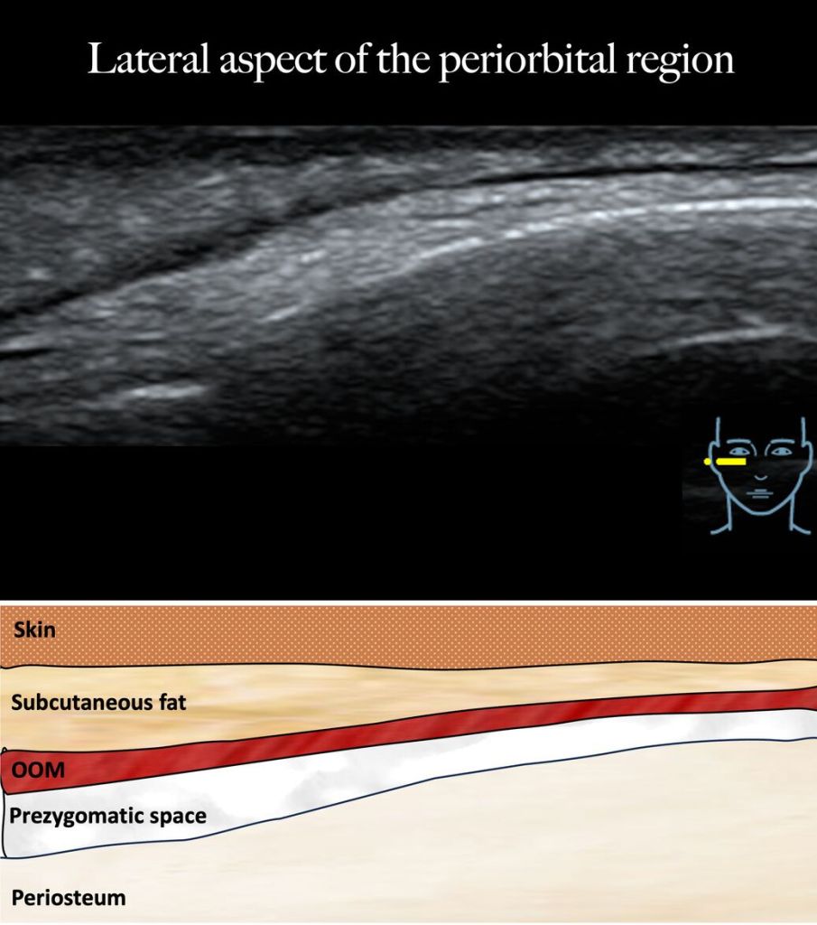
The treatment of the medial infraorbital region also termed the tear trough has become increasingly popular by the use of soft tissue fillers in a minimally invasive approach using a cannula.
A total of 246 tear troughs were injected and investigated originating from 123 study participants. The clinical outcome was evaluated 6 months after the treatment by independent observers based on standardized frontal images and the procedure was documented by ultrasound imaging.
On average, 0.26 (0.1) cc [range: 0.08–0.32] of soft tissue filler material was injected per tear trough. Tear trough depth was before the treatment rated as 2.12 (0.4), whereas after the treatment it was 1.15 (0.4) (p < 0.001). Hyperpigmentation score was 2.19 (0.4) before the treatment, whereas after the treatment it was 1.31 (0.5) (p < 0.001). Intraorbital fat pseudo-prolapse severity was rated before the treatment 1.88 (0.7), whereas it was rated after the treatment 1.14 (0.3) (p < 0.001). Wrinkle severity of the lower eyelid was rated before the treatment 1.51 (0.6), whereas it was rated after the treatment 1.12 (0.3) (p < 0.001).
The results of this retrospectively investigated case series revealed that the conducted injection technique for treating the tear trough for medial infraorbital hollowing with a cannula provided statistically significant clinical improvement with a limited adverse events profile. The technique utilized an injection approach which was perpendicularly oriented to the longitudinal axis of the tear trough thereby “bridging the gap instead of filling the entire valley.”
Bernardini, F., Skippen, B., Cetto, R., Calomeni, M., Cotofana, S., Urso, S., Paternostro, F. and Hartstein, M. (2024), Bridging the Gap Rather Than Filling the Entire Valley—Anatomic Insights When Treating the Medial Infraorbital Region. J Cosmet Dermatol. https://doi.org/10.1111/jocd.16582
Niccolò Fagni, Ferdinando Paternostro, Jacopo Junio Valerio Branca, Lorenzo Salerni, Marco Mandalà.
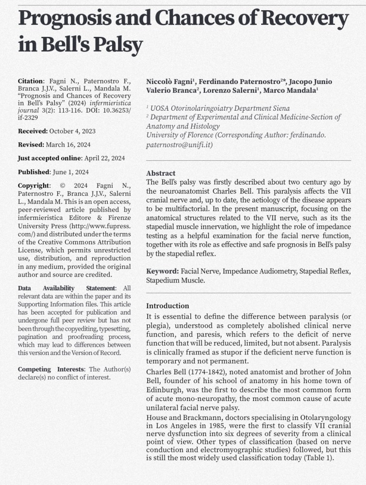
The Bell’s palsy was firstly described about two century ago by the neuroanatomist Charles Bell. This paralysis affects the VII cranial nerve and, up to date, the aetiology of the disease appears to be multifactorial.
In the present manuscript, focusing on the anatomical structures related to the VII nerve, such as its the stapedial muscle innervation, we highlight the role of impedance testing as a helpful examination for the facial nerve function, together with its role as effective and safe prognosis in Bell’s palsy by the stapedial reflex.
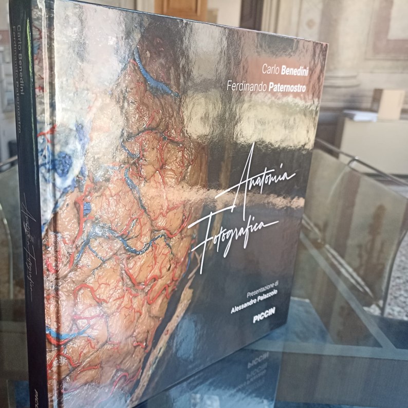
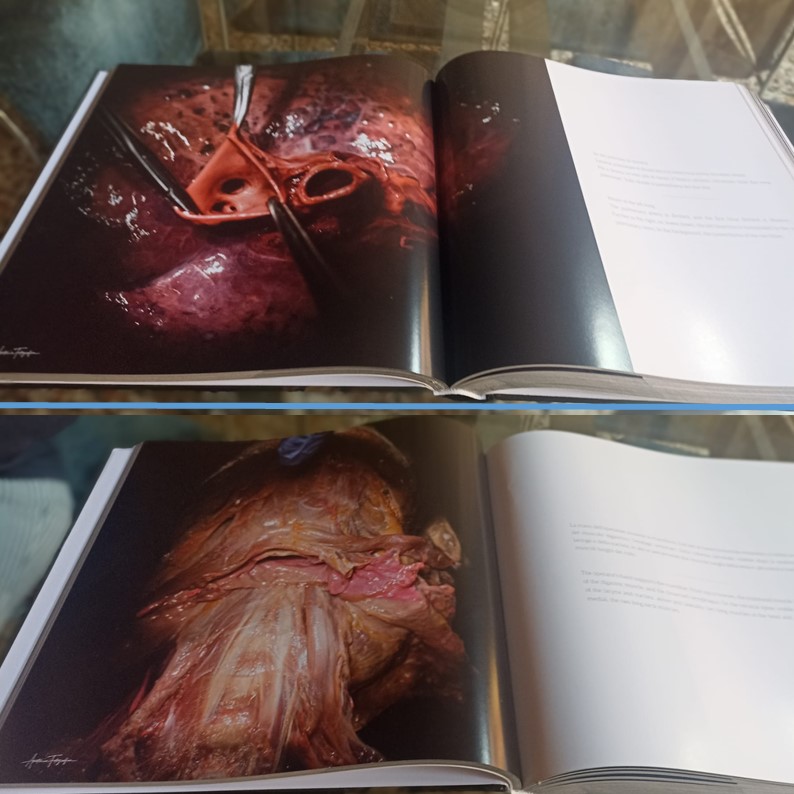
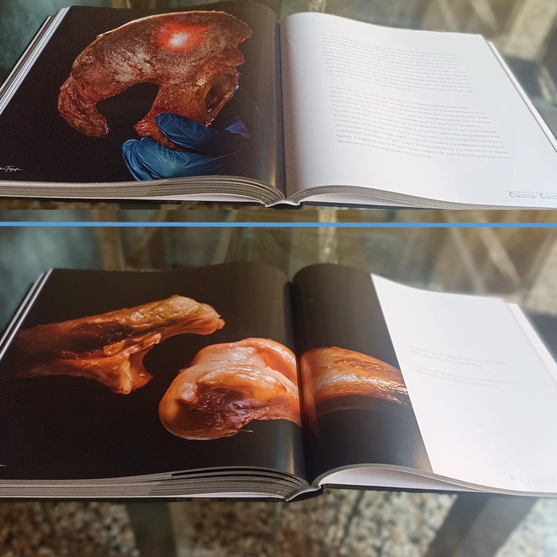
Le foto che potrete sfogliare in questo libro derivano da miei laboratori di Anatomia settoria (Anatomy Lab) realizzati a Verona pressoICLO, Teaching and Research Center. Qui è possibile studiare su preparati anatomici fresh frozen grazie al supporto della Nicola’s Foundation Onlus, che da sempre incentiva lo studio e la formazione in ambito medico scientifico.
Sono profondamente grato a tutto lo staff ICLO per la fondamentale ed efficiente disponibilità tecnica, amministrativa e logistica e al Dott. Gianni Sereni che, fin dal suo nascere e con lungimirante intuito, ha esaltato un progetto di formazione nazionale hands on, di alta qualità ma dai costi contenuti, poiché fondamentalmente rivolto a studenti universitari di indirizzo medico-sanitario.
Le immagini immortalate dal Dott. Carlo Benedini sono state realizzate, in questo ambito, con il supporto essenziale della dr.ssa Cristiana Veltro e del dott. Francesco Potenza competenti, appassionati e abili dissettori, che hanno condiviso con me l’esperienza delle prime edizioni di un progetto oggi fecondo anche grazie alla loro bravura e dedizione.
Durante le mie lezioni al tavolo cerco sempre di incrociare ed esplicitare ai discenti concetti di Anatomia palpatoria, topografica e sistematica con quanto il preparato “autonomamente” descrive. Molto spesso, poi, abbiamo la sorte di imbatterci in varianti anatomiche; tale esperienza è di grande valore per l’operatività nelle discipline chirurgiche, unitamente alla dimostrazione delle principali vie di accesso alle singole strutture e agli organi.
Le più moderne tecniche di imaging e le sofisticate metodiche operatorie illustrano con precisione e perizia tanti aspetti dell’Anatomia, ma non possono sostituire l’esperienza diretta sul cadavere, pratica antica ma fondamentale anche oggi nella formazione di studenti, specializzandi, specialisti.
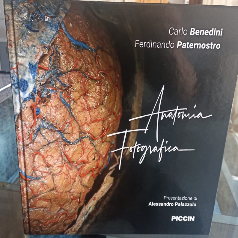
Carlo, egregio fotografo e appassionato anatomista, è riuscito con i suoi scatti a mescolare meraviglia, arte e rigore didattico, che ho provato a chiosare con le didascalie che completano le 516 pagine del libro.
Grazie al Prof. Alessandro Palazzolo per la preziosa e affettuosa presentazione e per gli incoraggiamenti che non sono mai mancati in corso d’opera. Grazie al Dott. Nicola Piccin che ha creduto nella originalità del nostro lavoro e a tutti coloro che hanno seguito il non semplice iter della realizzazione tipografica.
Felice, orgoglioso e grato per aver realizzato, con Carlo, questo esclusivo e originale volume.
Experimental and Clinical Medicine, University of Firenze, Firenze, ITA
Physical Medicine and Rehabilitation, ICLO Teaching and Research Center, Verona, ITA
Otorinolaringoiatry, Azienda Ospedaliero-Universitaria Senese (UOSA), Siena, ITA
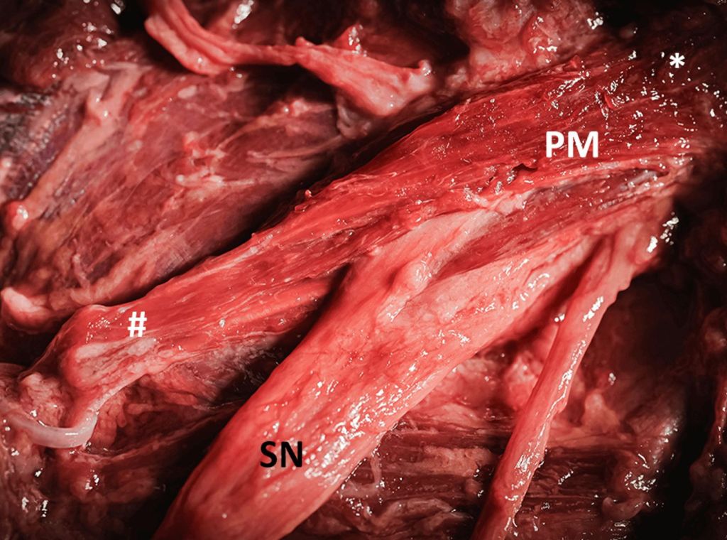
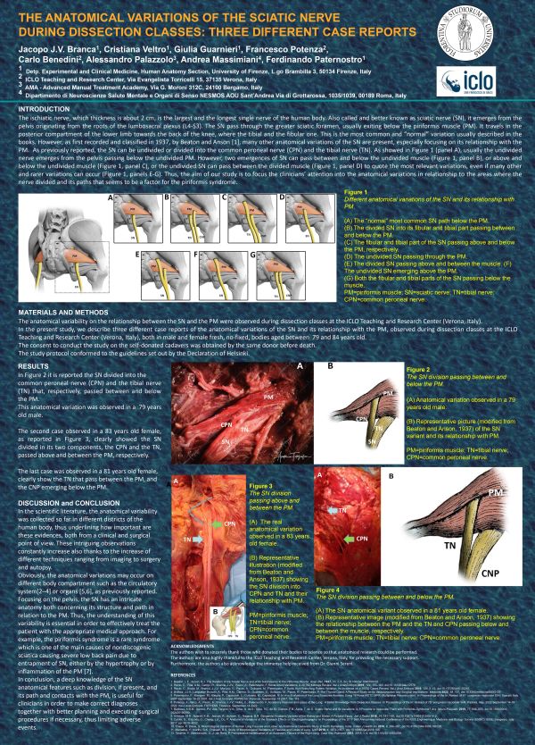
Knowledge of anatomical variability is extremely important in order to better understand the etiology of pain, if present, or to avoid iatrogenic consequences. Sometimes the anatomical “anomalies” have the same anamnesis but different causes. For example, sciatic neuralgia may be caused by a herniated disc or it may have a different origin. The sciatic nerve (SN), also known as the ischial nerve, is the widest in the human body. This huge peripheral nerve originates from the roots of the lumbosacral plexus (L4-S3) and passes through the great sciatic foramen, under the piriformis muscle (PM). However, there is much variability in the pattern of SNs about the muscle, which has been known since the first half of the 20th century. In the present study, we describe six different case reports of anatomical variations of the SN and its interplay with the PM. The observations were made during dissection classes at the ICLO Teaching and Research Centre (Verona, Italy), on both male and female cadavers aged between 58 and 84 years. The SN was reported as a single and divided nerve into the tibial nerve (TN) and the common peroneal nerve (CPN), passing alone above, below, or between the PM. However, the two parts of the SN may also interact with the PM in different ways, adding to the anatomical variability. A thorough knowledge of the anatomical variations in any part of the human body is extremely important. The various techniques used, from imaging to autopsy or surgery, are also useful in the SN pathway. Thus, the anatomical features and the understanding of each variation are useful for a correct approach that can lead to an effective and correct treatment with a favorable outcome.
Acknowledgements We are extremely grateful to Dr. Alessandro Palazzolo, Dr. Daniele Pignatelli, Dr. Cristiana Veltro, Anna Venzi, and Aurora Baroni for their skills and dedication demonstrated during the dissection classes performed together with the ICLO staff and Prof. Ferdinando Paternostro.
“Sembra che tu abbia condiviso o inviato un video (è una foto, in realtà..) che mostra contenuti di violenza esplicita.”
Da anni sto scrivendo post (oltre 800) che spiegano la meravigliosa costruzione del Corpo Umano, con rispetto, stupore e con la voglia di condividere conoscenza…invece secondo gli algoritmi di Instagram, sotto sotto desidero istigare alla violenza e per di più in maniera chiara ed esplicita.
Non ho parole. Sono profondamente amareggiato e deluso.
Non ho bisogno di spiegare ad un “umano” cosa sta facendo un Professore di Anatomia nella foto incriminata (nel contesto di un post in cui l’occasione didattica era più che chiara)…Con le macchine con cui mi sono interfacciato fino ad ora mi è risultato impossibile!
Condividete la pagina, per favore, condividete un post. Forse riusciremo a convincere le intelligenze naturali e artificiali che l’Anatomia (per Tutti) è cosa seria, importante e apprezzata.
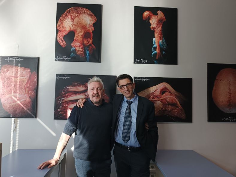
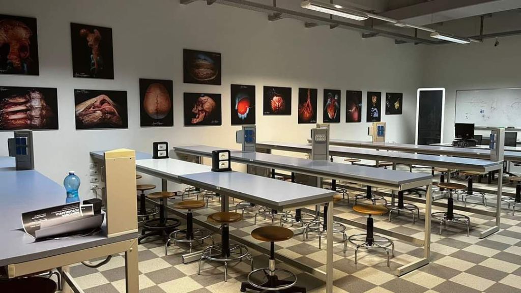
Lo scorso 19 gennaio sono stato ospite del Dipartimento di Medicina dell’Università Insubria di Varese. L’occasione era l’inaugurazione di una mostra permanente realizzata con una serie di splendide fotografie dell’AmicoDott. Carlo Benedini. Carlo, mosso da comune passione per l’Anatomia, ha avuto la pazienza di seguirmi in numerose dissezioni svolte presso ICLO, Verona (Anatomy Lab), durante le quali ha realizzato un numero infinito di bellissimi scatti. Da questa immensa mole di immagini ha distillato quelle più significative, che sono diventate prima la colonna portante di una pagina Instagram di successo (Anatomia Fotografica), la mostra di Varese e infine un volume di oltre duecentoquaranta tavole commentate che uscirà, con la prefazione del Prof. Alessandro Palazzolo, per i tipi di Piccin.
Voglio di cuore ringraziare la Prof.ssa Marina Protasoni, appassionata Docente e fine Anatomista, che ha voluto fortemente l’esposizione e ha organizzato la giornata di presentazione della stessa.
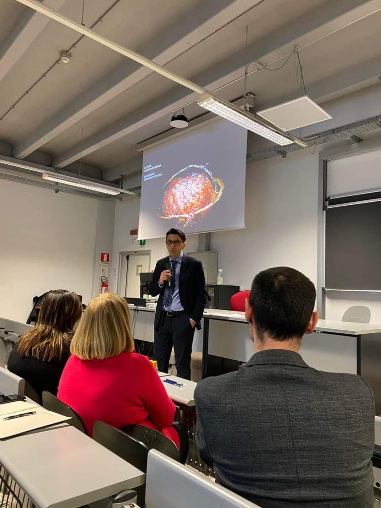
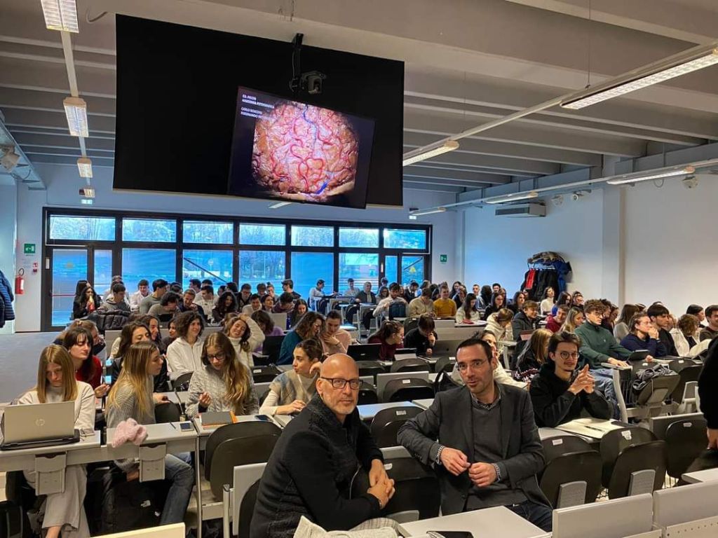
Grazie al Prof. Giulio Carcano, Direttore DIMIT (Dipartimento di Medicina e Innovazione Tecnologica) e al Prof. Alberto Passi, Presidente della Scuola di Medicina, ai Colleghi presenti, Prof. ssa Marcella Reguzzoni, il Prof. Pier Antonio Zecca. Grazie ai numerosi, attenti e partecipi Studenti del Corso di Laurea in Medicina e Chirurgia, ai quali ho raccontato di come l’evoluzione ha modellato il nostro corpo e in particolare Sistema Nervoso Centrale. Il titolo della relazione era “L’uomo è un animale addomesticato?” La risposta è “sì…” ma se volete sapere da chi venite a scoprirlo ai prossimi appuntamenti con Anatomia per Tutti!
Kamal Ivory, Rossella Angotti, Mario Messina, Denise Bonente, Ferdinando Paternostro, Massimo Gulisano and Claudio Nicoletti
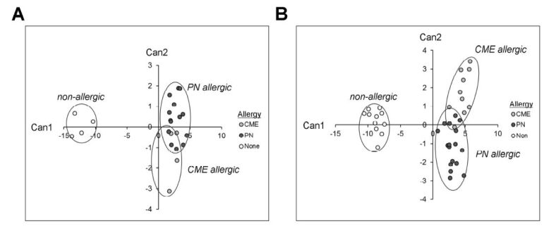
Ricerca sulle alterazioni del sistema immunitario di bambini affetti da allergie alimentari. Lo studio evidenzia che in soggetti allergici alcuni tipi cellulari importanti per le risposte immunitarie riescono a sopravvivere più a lungo evitando il fenomeno dell’apoptosi o morte cellulare. Questo grazie all’espressione del gene Bcl-2 che blocca l’apoptosi. La resistenza all’apoptosi è alla base di molte malattie; per la prima volta questo difetto viene associato anche alle reazioni allergiche.
All allergic responses to food indicate the failure of immunological tolerance, but it is unclear why cow’s milk and egg (CME) allergies resolve more readily than reactivity to peanuts (PN). We sought to identify differences between PN and CME allergies through constitutive immune status and responses to cognate and non-cognate food antigens. Children with confirmed allergy to CME (n = 6) and PN (n = 18) and non-allergic (NA) (n = 8) controls were studied. Constitutive secretion of cytokines was tested in plasma and unstimulated mononuclear cell (PBMNC) cultures. Blood dendritic cell (DC) subsets were analyzed alongside changes in phenotypes and soluble molecules in allergen-stimulated MNC cultures with or without cytokine neutralization. We observed that in allergic children, constitutively high plasma levels IL-1, IL-2, IL-4, IL-5 and IL-10 but less IL-12p70 than in non-allergic children was accompanied by the spontaneous secretion of sCD23, IL-1, IL-2, IL-4, IL-5, IL-10, IL-12p70, IFN-and TNF- in MNC cultures. Furthermore, blood DC subset counts differed in food allergy. Antigen-presenting cell phenotypic abnormalities were accompanied by higher B and T cell percentages with more Bcl-2 within CD69+ subsets. Cells were generally refractory to antigenic stimulation in vitro, but IL-4 neutralization led to CD152 downregulation by CD4+ T cells from PN allergic children responding to PN allergens. Canonical discriminant analyses segregated non-allergic and allergic children by their cytokine secretion patterns, revealing differences and areas of overlap between PN and CME allergies. Despite an absence of recent allergen exposure, indication of in vivo activation, in vitro responses independent of challenging antigen and the presence of unusual costimulatory molecules suggest dysregulated immunity in food allergy. Most importantly, higher Bcl-2 content within key effector cells implies survival advantage with the potential to mount abnormal responses that may give rise to the manifestations of allergy. Here, we put forward the hypothesis that the lack of apoptosis of key immune cell types might be central to the development of food allergic reactions.
In Anatomia parliamo di “variante” ogni volta che una struttura ha una morfologia che si discosta da quella osservata nella maggior parte degli individui e rappresenta una deviazione dagli standard (la norma nella sua accezione statistica) condivisi nei libri di testo e insegnati nelle aule universitarie.
Le varianti, tuttavia, non inficiano la funzionalità dell’organo che “modificano” e per questo rientrano in un quadro di normalità (in questo caso intendendo un appropriato funzionamento), al contrario delle anomalie (congenite o meno) che già nella definizione evidenziano il loro aspetto patologico.
Conformazioni anatomiche particolari, tuttavia, possono interferire con procedure diagnostiche e aumentare i rischi di specifici atti chirurgici. Per questo la conoscenza e lo studio delle variazioni anatomiche dalla norma è un presupposto indispensabile per la pratica medica.
In sintesi (e per punti) lo studio delle varianti anatomiche è importante per diverse ragioni:
La comprensione della normalità: Lo studio delle varianti aiuta a definire meglio ciò che è considerato “normale” all’interno della diversità anatomica umana. Ciò consente di riconoscere quando una caratteristica è effettivamente una variante innocua anziché una condizione patologica.
La diagnosi e il trattamento: Alcune varianti anatomiche possono essere associate a condizioni mediche o sintomi. La conoscenza di queste varianti è essenziale per una diagnosi accurata e per determinare il trattamento più appropriato.
La prevenzione e gestione delle complicanze: La consapevolezza della presenza delle varianti può aiutare a prevenire complicanze durante procedure mediche o chirurgiche. Ad esempio, durante un intervento chirurgico, conoscere la posizione di un’arteria variante può evitare danni accidentali.
La ricerca: Lo studio delle varianti anatomiche può aiutare a comprendere meglio l’evoluzione dell’anatomia umana e le cause genetiche di alcune modificazioni. Questo può portare a scoperte significative e all’identificazione di nuovi bersagli per terapie e trattamenti.
L’educazione medica: L’inclusione delle varianti anatomiche nei programmi di formazione medica è essenziale per preparare i futuri medici a riconoscere e gestire una vasta gamma di condizioni cliniche.
La consulenza genetica: Le varianti anatomiche possono essere ereditate geneticamente. Lo studio delle varianti anatomiche è pertanto rilevante per la consulenza genetica.
La promozione della diversità: Riconoscere e accettare la diversità anatomica umana promuove una migliore comprensione e tolleranza delle differenze tra le persone. Questo, in ultima analisi è un contributo ulteriore alla promozione dell’uguaglianza e dell’inclusione.
L’attività settoria, che con l’imaging e la chirurgia consente di individuare e documentare eventuali varianti rispetto alla normale Anatomia umana, costituisce, anche per questo, un supporto fondamentale per la Medicina.
L’ulltima ricerca in questo filone di investigazione, riguarda le variazioni morfologiche e topografiche del nervo sciatico in particolare in relazione al muscolo piriforme.
Ne abbiamo parlato prima al 76° Congresso Nazionale della Società Italiana di Anatomia e Istologia (Modena, 11-13 settembre 2023). Per scaricare il poster in formato pdf, stampabile, clicca qui.
Jacopo J.V. Branca, Cristiana Veltro, Giulia Guarnieri, Francesco Potenza, Carlo Benedini, Alessandro Palazzolo, Andrea Massimiani, Ferdinando Paternostro The anatomical variations of the sciatic nerve during dissection classes: three different case reports Italian Journal of Anatomy and Embryology 121(1) Supplement: 267, 2023 Supplement Firenze University Press ISSN 1122-6714 (print) | ISSN 2038-5129 (online).
Successivamente, con il rinvenimento di numerose altre varianti, abbiamo pubblicato questo articolo su Cureus: