Nel 2013 attivai quasi per celia una pagina Facebook con questo nome perché, ricevendo costantemente da tanti Studenti richieste di spiegazioni o chiarimenti sui più disparati argomenti di Anatomia Umana, pensavo (e oggi ne sono ancora più convinto) di condividere con tutti le risposte, gli approfondimenti e, soprattutto, la passione per questa fondamentale disciplina biomedica.
Molti considerano l’Anatomia una materia preclinica e infatti all’Università viene studiata sempre nei primi anni di corso. Personalmente la ritengo “trans-clinica”, perché permea di sé tutto il sapere medico-scientifico. Lo permea in maniera “spirale”, ovvero con sempre maggiore capacità e necessità di approfondimento, durante la formazione prima e la carriera poi di tutto il personale Sanitario.
L’Anatomia è lo studio delle strutture di un organismo e dei rapporti tra le sue parti. La parola deriva dal greco e vuol dire “per mezzo della dissezione”, che per secoli è stata la sola tecnica utilizzata per isolare e studiare le singole parti del corpo umano. Ma la dissezione, per quanto fondamentale, non è l’unica chiave di lettura di questa disciplina.
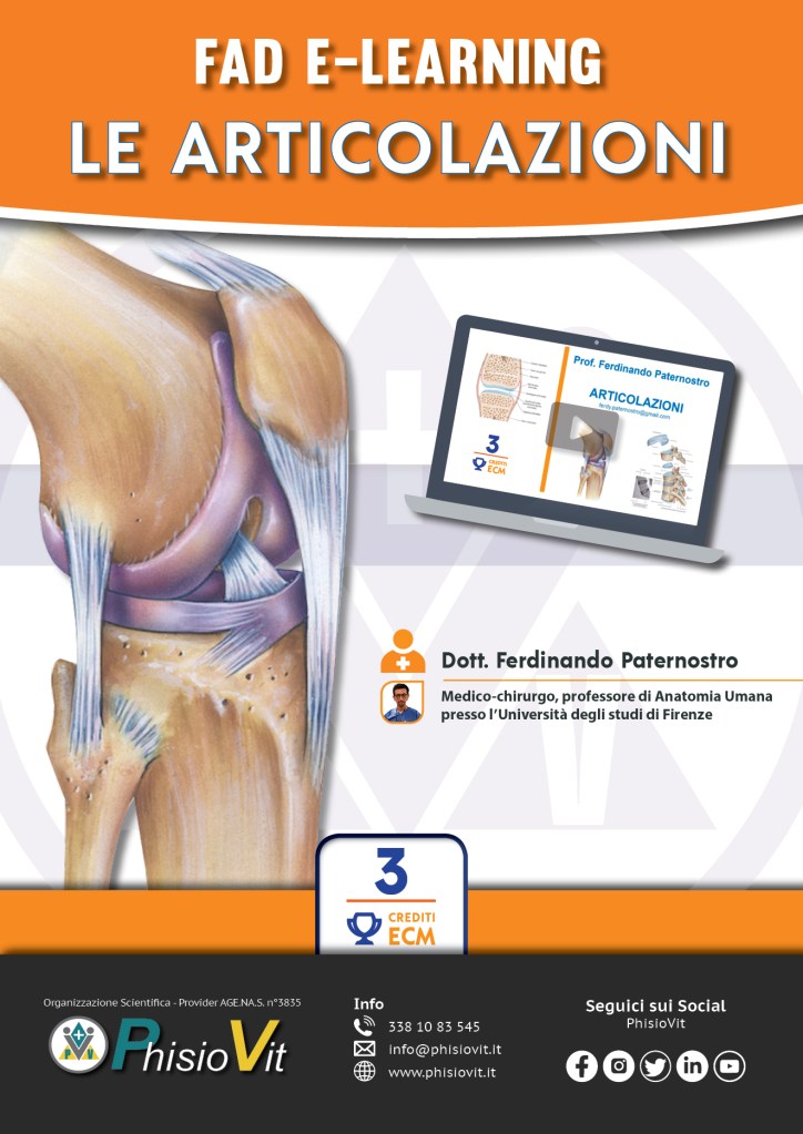
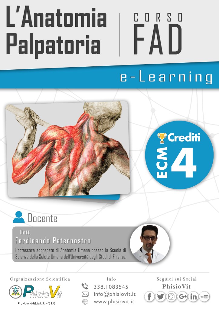
Infatti oltre all’Anatomia macroscopica, che studia strutture sufficientemente grandi da essere osservabili ad occhio nudo, c’è l’Anatomia di superficie, che valuta la forma generale ed esterna delle varie aree anatomiche e riconosce le strutture attraverso le tecniche palpatorie, l’Anatomia topografica o regionale, chesi occupa degli organi in relazione alla loro posizione nel corpo e ai loro reciproci rapporti, l’Anatomia sistematica, che scompone l’organismo in sistemi e apparati e, nell’ambito di essi, studia ciascun organo. Dalla sistematica, in genere, si parte per un primo approccio alla materia.
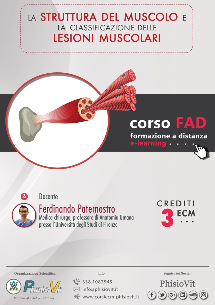
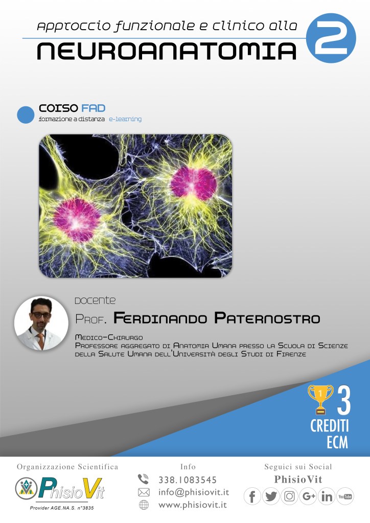
Attraverso l’Anatomia microscopica si valutano le strutture non visibili ad occhio nudo, che spesso consentono di riconoscere e distinguere le diverse porzioni dello stesso organo (parlare di Anatomia microscopica-topografica sembra un ossimoro … ma a pensarci bene non lo è !).
A questo livello si apprezza la “consustanziazione” della Microscopica con le discipline sorelle dell’Istologia (lo studio dei tessuti) e della Citologia (lo studio della cellula).
L’Anatomia dello sviluppo di occupa delle modificazioni morfologiche che avvengono tra il concepimento e la maturazione fisica. La disciplina che studia i processi dello sviluppo è l’Embriologia. L’ Anatomia radiologica si focalizza sulla nomenclatura e sull’aspetto delle singole parti del corpo umano così come appaiono alla radioscopia, alla radiografia, all’ecografia e alle più recenti tecniche di diagnostica per immagini (TC, RMN, PET). L’Anatomia chirurgica studia i problemi anatomici relativi a malattie che ottengono risoluzione con metodi chirurgici, ai loro sintomi e agli interventi corrispondenti. L’Anatomia clinica segue le modificazioni anatomiche durante lo sviluppo delle patologie. L’Anatomia comparata evidenzia similitudini e differenze di forma e di struttura in organismi diversi fra loro; appartiene alle Scienze Naturali.
ANATOMIA PER TUTTI è una Community virtuale di Studiosi, Studenti e appassionati di Anatomia Umana. Non è più soltanto una pagina Facebook, ma anche un canale ricco di contenuti su Youtube, una pagina su Instagram e un canale Telegram. Ha anche tanti e qualificati redattori che con me condividono la passione della comunicazione scientifica e dell’insegnamento.
Con sempre nuovi contenuti, ogni giorno sul web studiamo e approfondiamo insieme le bellezze del Corpo, lo strumento più prezioso che abbiamo, da tutelare, conoscere e far crescere in salute.
Nel nostro Corpo quotidianamente ogni singola cellula racconta la storia dell’evoluzione, ogni Organo l’armonia della vita, ogni Apparato il valore delle sinergie. La nostra Community è il luogo virtuale dove condividere sapere e meraviglia per l’incommensurabile bellezza dell’Anatomia umana.
FACEBOOK
https://www.facebook.com/anatomiapertutti/
YOUTUBE
https://www.youtube.com/channel/UC0UgcnjTvobPMeX0P78oUsw
INSTAGRAM
https://www.instagram.com/anatomiapertutti/
TELEGRAM
https://t.me/pertuttianatomia